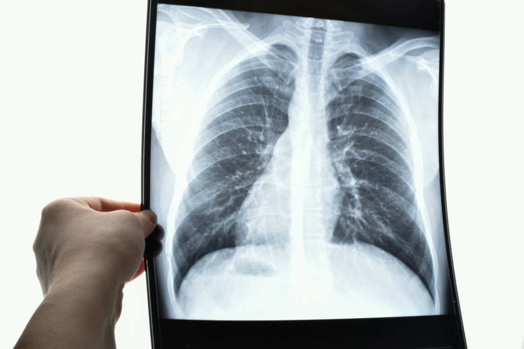
So, you’ve been experiencing some discomfort in your chest and upper abdomen, and your doctor suspects it might be a hiatal hernia. You’re probably wondering how they’re going to confirm this diagnosis. Well, let me fill you in on the important role that X-ray plays in the diagnosis of hiatal hernias. By using X-ray imaging, doctors are able to get a clear picture of the hiatal area and identify any abnormalities or protrusions. This non-invasive and relatively simple procedure provides valuable information that helps in determining the best course of treatment for you. So, let’s take a closer look at how X-ray plays a crucial role in diagnosing hiatal hernias.

What is a Hiatal Hernia?
A hiatal hernia occurs when a part of the stomach pushes through the diaphragm and into the chest cavity. The diaphragm normally has a small opening called the hiatus, which allows the esophagus to pass through and connect to the stomach. However, in some cases, the stomach can herniate through this opening, leading to a hiatal hernia.
Types of Hiatal Hernias
There are two main types of hiatal hernias: sliding hiatal hernia and paraesophageal hiatal hernia.
Sliding Hiatal Hernia: This is the most common type of hiatal hernia. In a sliding hernia, the stomach and the esophagus slide into the chest together. The junction between the stomach and the esophagus, known as the gastroesophageal junction, moves upwards.
Paraesophageal Hiatal Hernia: In this type of hiatal hernia, a portion of the stomach pushes through the hiatus and lies alongside the esophagus. Unlike sliding hernias, the gastroesophageal junction remains in its normal position.
Importance of Hiatal Hernia Diagnosis
Early detection of a hiatal hernia is crucial as it allows for prompt management of symptoms and prevents the development of complications.
Early Detection
Diagnosing a hiatal hernia at an early stage allows healthcare professionals to provide appropriate treatment and prevent the condition from worsening. Early intervention can help alleviate symptoms and improve the overall quality of life for individuals with hiatal hernias.
Managing Symptoms
Diagnosis enables healthcare professionals to develop a personalized plan to manage the symptoms associated with hiatal hernias. By understanding the specific type and severity of the hernia, appropriate lifestyle modifications and medical interventions can be implemented to reduce discomfort and enhance overall well-being.
Prevention of Complications
Early diagnosis of hiatal hernias also helps in identifying any potential complications that may arise. Prompt intervention can prevent complications such as gastroesophageal reflux disease (GERD), esophagitis, and Barrett’s esophagus. By addressing the hernia early, healthcare professionals can minimize the risk of further damage to the esophagus and stomach.
Role of X-ray in Hiatal Hernia Diagnosis
X-ray, also known as radiography, plays a vital role in diagnosing hiatal hernias. It is a non-invasive imaging technique that allows healthcare professionals to visualize the internal structures of the chest and abdomen.
Overview
During an X-ray examination, a small amount of radiation is used to create images of the diaphragm, stomach, and esophagus. These images help in identifying and evaluating hiatal hernias, as well as assessing their size and severity.
Procedure
The X-ray procedure for hiatal hernia diagnosis involves positioning the patient, taking X-ray images, and interpreting the results. It is a relatively quick and straightforward process that is usually performed in a radiology department or clinic.
Preparation
Prior to the X-ray examination, certain preparations may be necessary. These may include fasting for a specific period, discontinuing certain medications, and removing any metal objects that may interfere with the imaging process. It is important to follow these preparation guidelines provided by the healthcare provider to ensure accurate results.
Benefits of X-ray for Hiatal Hernia Diagnosis
X-ray offers several advantages when it comes to diagnosing hiatal hernias. Understanding these benefits can help individuals and healthcare professionals make informed decisions about the most appropriate diagnostic approach.
Non-invasive Imaging
One of the key benefits of X-ray is that it is a non-invasive imaging modality. This means that there is no need for any incisions or invasive procedures to visualize the hernia. As a result, there is minimal discomfort and a lower risk of complications compared to more invasive diagnostic methods.
Rapid Results
X-ray examinations typically provide immediate results. The X-ray images are captured in real-time, allowing the radiologist to interpret them without delay. This allows for a quick diagnosis and enables timely intervention if necessary. Rapid results can significantly reduce anxiety and uncertainty for individuals awaiting a diagnosis.
Signs and Symptoms of Hiatal Hernia
Hiatal hernias can present with a variety of signs and symptoms, which can vary in severity from individual to individual. It is important to be aware of these symptoms to seek appropriate medical attention if they arise.
Heartburn
Heartburn is one of the most common symptoms associated with hiatal hernias. It is characterized by a burning sensation in the chest, often accompanied by a sour or acidic taste in the mouth. The discomfort usually occurs after meals or when lying down and can be exacerbated by certain foods and behaviors.
Chest Pain
Chest pain can occur in individuals with hiatal hernias, although it is important to differentiate it from other serious cardiac conditions. The pain may be dull or sharp and can sometimes radiate to the arms, neck, or back. It is important to seek medical attention if chest pain persists or is accompanied by other concerning symptoms.
Difficulty Swallowing
Hiatal hernias can cause difficulty swallowing, also known as dysphagia. Individuals may feel as though food is getting stuck in their throat or chest, leading to discomfort and a feeling of fullness. Dysphagia can significantly impact a person’s ability to eat and may require dietary adjustments or medical interventions.
Regurgitation
Regurgitation refers to the backflow of stomach contents into the esophagus or mouth. It can be experienced as a sour or bitter taste, accompanied by a sensation of fluid or food coming back up. Regurgitation can be distressing and may occur after meals or when lying down.
Nausea
Nausea is another symptom that can be associated with hiatal hernias. It may be experienced as a general feeling of queasiness or the urge to vomit. Nausea can be triggered by the hernia itself or as a result of associated conditions such as GERD.
Other Diagnostic Methods for Hiatal Hernia
While X-ray is an effective diagnostic tool for hiatal hernias, there are other methods available to confirm the presence of a hernia and assess its severity.
Endoscopy
Endoscopy involves the use of a flexible tube with a camera and light source, known as an endoscope, to visualize the esophagus, stomach, and duodenum. It allows for direct visualization of the hernia and provides detailed information about the condition of the esophageal lining. Endoscopy is often performed if a hiatal hernia is suspected but not clearly seen on X-ray.
Esophageal Manometry
Esophageal manometry measures the muscle contractions in the esophagus and assesses the function of the lower esophageal sphincter. It can provide valuable information about the pressure changes during swallowing and help identify abnormalities that may contribute to the development of hiatal hernias.
Barium Swallow Test
A barium swallow test involves swallowing a liquid contrast agent that contains barium. X-ray images are then taken as the barium flows through the esophagus and stomach. This test helps visualize any abnormalities, including hiatal hernias, and provides detailed information about the shape and function of the esophagus.
Preparing for an X-ray Examination
Before undergoing an X-ray for hiatal hernia diagnosis, certain instructions and guidelines need to be followed to ensure accurate results.
Fasting Instructions
In most cases, individuals will be asked to fast for a specific period before the X-ray examination. This typically involves abstaining from food and drink for a set amount of time to ensure that the stomach is empty during the procedure. Fasting instructions should be provided by the healthcare provider, and it is crucial to follow them closely for accurate imaging.
Medication Guidelines
Healthcare professionals may provide specific guidelines regarding medication use before the X-ray examination. Certain medications, particularly those that affect the digestive system, may need to be discontinued temporarily to ensure accurate evaluation of the hernia. It is essential to consult with the healthcare provider and follow their instructions regarding medication use.
Removing Metal Objects
Metal objects, such as jewelry or clothing accessories, may interfere with the X-ray imaging process. As a result, it is important to remove any metal objects from the chest and abdominal area before the examination. This helps ensure clear and unobstructed images of the hernia and surrounding structures.
Procedure of X-ray for Hiatal Hernia Diagnosis
The X-ray procedure for hiatal hernia diagnosis involves several steps, including positioning the patient, taking X-ray images, and interpreting the results.
Positioning the Patient
The patient will be positioned in various ways to obtain optimal images of the hernia. This may include lying on the X-ray table in different positions or standing against the X-ray machine. The radiology technician will guide the patient through the positioning process to capture the necessary images.
Taking X-ray Images
Once the patient is positioned correctly, the radiology technician will operate the X-ray machine to capture images of the chest and abdominal area. The patient will be instructed to hold their breath for a few seconds while the images are taken to ensure clear and sharp results. Multiple images may be taken from different angles to provide a comprehensive view of the hernia.
Radiologist Interpretation
After the X-ray images are taken, they will be sent to a radiologist for interpretation. The radiologist will carefully review the images and assess for the presence of a hiatal hernia, its size, and any associated complications. The radiologist’s findings will be documented and shared with the healthcare provider, who will communicate the results to the patient.
Interpreting X-ray Results
The radiologist’s interpretation of the X-ray results is crucial in determining the presence, size, and severity of a hiatal hernia. This information helps guide further management and treatment decisions.
Identification of Hiatal Hernia
The X-ray images allow the radiologist to identify the presence of a hiatal hernia. By evaluating the position of the stomach in relation to the diaphragm and esophagus, the radiologist can confirm the diagnosis and determine the specific type of hernia.
Assessment of Size and Severity
X-ray images provide important information about the size and severity of a hiatal hernia. The radiologist can measure the extent of the herniation and assess whether it is causing any compression or displacement of surrounding structures. This information helps determine the appropriate treatment approach and guides discussions with the patient.
Identifying Complications
In addition to diagnosing the hiatal hernia itself, X-ray can sometimes reveal complications or associated conditions. The radiologist may identify signs of gastroesophageal reflux, esophagitis, or other abnormalities during the interpretation. This information is valuable in developing a comprehensive treatment plan and managing any additional concerns.
Limitations and Considerations of X-ray
While X-ray is an invaluable tool in diagnosing hiatal hernias, it does have certain limitations and considerations that should be taken into account.
False Negatives and Positives
X-ray images may not always clearly show the presence of a small or subtle hiatal hernia. In some cases, a hernia may be missed on X-ray, resulting in a false negative. Conversely, a hiatal hernia may appear on X-ray images when one is not actually present, leading to a false positive. These considerations highlight the importance of combining X-ray findings with other diagnostic methods to achieve an accurate diagnosis.
Exposure to Radiation
X-ray imaging involves exposure to a small amount of radiation. While the dose of radiation used during an X-ray examination is generally considered safe, individuals who require frequent or repeated X-rays for monitoring purposes should discuss the potential risks with their healthcare provider.
Other Diagnostic Modalities
While X-ray is a valuable tool for hiatal hernia diagnosis, there are other diagnostic modalities available that may provide additional information. Endoscopy, esophageal manometry, and barium swallow tests can all complement X-ray findings and provide a more comprehensive assessment of the hiatal hernia and its associated complications.
In conclusion, X-ray plays a pivotal role in the diagnosis of hiatal hernias. By providing non-invasive and rapid results, it enables early detection, symptom management, and prevention of complications. While X-ray has its limitations, its benefits and utility in hiatal hernia diagnosis cannot be overstated. By understanding the signs and symptoms of hiatal hernias, individuals can seek timely medical attention and access the appropriate diagnostic methods, ensuring comprehensive care and improved quality of life.
